You are browsing environment: HUMAN GUT
CAZyme Information: MGYG000000754_00548
You are here: Home > Sequence: MGYG000000754_00548
Basic Information |
Genomic context |
Full Sequence |
Enzyme annotations |
CAZy signature domains |
CDD domains |
CAZyme hits |
PDB hits |
Swiss-Prot hits |
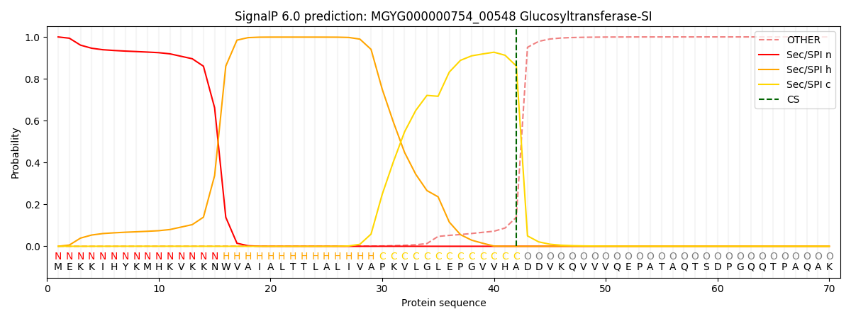
SignalP and Lipop annotations |
TMHMM annotations
Basic Information help
| Species | Streptococcus sp900555155 | |||||||||||
|---|---|---|---|---|---|---|---|---|---|---|---|---|
| Lineage | Bacteria; Firmicutes; Bacilli; Lactobacillales; Streptococcaceae; Streptococcus; Streptococcus sp900555155 | |||||||||||
| CAZyme ID | MGYG000000754_00548 | |||||||||||
| CAZy Family | GH70 | |||||||||||
| CAZyme Description | Glucosyltransferase-SI | |||||||||||
| CAZyme Property |
|
|||||||||||
| Genome Property |
|
|||||||||||
| Gene Location | Start: 1360; End: 4398 Strand: - | |||||||||||
CAZyme Signature Domains help
| Family | Start | End | Evalue | family coverage |
|---|---|---|---|---|
| GH70 | 332 | 983 | 0 | 0.7982565379825654 |
CDD Domains download full data without filtering help
| Cdd ID | Domain | E-Value | qStart | qEnd | sStart | sEnd | Domain Description |
|---|---|---|---|---|---|---|---|
| pfam02324 | Glyco_hydro_70 | 0.0 | 332 | 982 | 1 | 638 | Glycosyl hydrolase family 70. Members of this family belong to glycosyl hydrolase family 70 Glucosyltransferases or sucrose 6-glycosyl transferases (GTF-S) catalyze the transfer of D-glucopyramnosyl units from sucrose onto acceptor molecules, EC:2.4.1.5. This family roughly corresponds to the N-terminal catalytic domain of the enzyme. Members of this family also contain the Putative cell wall binding domain pfam01473, which corresponds with the C-terminal glucan-binding domain. |
| PRK09441 | PRK09441 | 2.78e-08 | 461 | 812 | 188 | 448 | cytoplasmic alpha-amylase; Reviewed |
| cd11318 | AmyAc_bac_fung_AmyA | 2.27e-06 | 461 | 725 | 186 | 389 | Alpha amylase catalytic domain found in bacterial and fungal Alpha amylases (also called 1,4-alpha-D-glucan-4-glucanohydrolase). AmyA (EC 3.2.1.1) catalyzes the hydrolysis of alpha-(1,4) glycosidic linkages of glycogen, starch, related polysaccharides, and some oligosaccharides. This group includes bacterial and fungal proteins. The Alpha-amylase family comprises the largest family of glycoside hydrolases (GH), with the majority of enzymes acting on starch, glycogen, and related oligo- and polysaccharides. These proteins catalyze the transformation of alpha-1,4 and alpha-1,6 glucosidic linkages with retention of the anomeric center. The protein is described as having 3 domains: A, B, C. A is a (beta/alpha) 8-barrel; B is a loop between the beta 3 strand and alpha 3 helix of A; C is the C-terminal extension characterized by a Greek key. The majority of the enzymes have an active site cleft found between domains A and B where a triad of catalytic residues (Asp, Glu and Asp) performs catalysis. Other members of this family have lost the catalytic activity as in the case of the human 4F2hc, or only have 2 residues that serve as the catalytic nucleophile and the acid/base, such as Thermus A4 beta-galactosidase with 2 Glu residues (GH42) and human alpha-galactosidase with 2 Asp residues (GH31). The family members are quite extensive and include: alpha amylase, maltosyltransferase, cyclodextrin glycotransferase, maltogenic amylase, neopullulanase, isoamylase, 1,4-alpha-D-glucan maltotetrahydrolase, 4-alpha-glucotransferase, oligo-1,6-glucosidase, amylosucrase, sucrose phosphorylase, and amylomaltase. |
| TIGR03715 | KxYKxGKxW | 7.77e-06 | 4 | 26 | 1 | 23 | KxYKxGKxW signal peptide. This model describes a novel form of signal peptide that occurs as an N-terminal domain with a recognizable motif, reminiscent of the YSIRK and PEP-CTERM forms of signal peptide. This domain tends to occur on long, low-complexity (usually Serine-rich and heavily glycosylated) proteins of the Firmicutes, and (as with YSIRK) the majority of these proteins have the LPXTG cell wall-anchoring motif at the C-terminus. |
| pfam19127 | Choline_bind_3 | 4.10e-05 | 226 | 264 | 5 | 44 | Choline-binding repeat. Pair of presumed choline-binding repeats often found adjacent to pfam01473. |
CAZyme Hits help
| Hit ID | E-Value | Query Start | Query End | Hit Start | Hit End |
|---|---|---|---|---|---|
| QQK99641.1 | 0.0 | 1 | 983 | 2 | 984 |
| QRO07363.1 | 0.0 | 1 | 983 | 2 | 984 |
| VEF78659.1 | 0.0 | 1 | 983 | 1 | 983 |
| QLL96091.1 | 0.0 | 1 | 983 | 2 | 984 |
| BAA95201.1 | 0.0 | 1 | 983 | 2 | 984 |
PDB Hits download full data without filtering help
| Hit ID | E-Value | Query Start | Query End | Hit Start | Hit End | Description |
|---|---|---|---|---|---|---|
| 3AIB_A | 2.49e-270 | 282 | 982 | 2 | 691 | CrystalStructure of Glucansucrase [Streptococcus mutans],3AIB_B Crystal Structure of Glucansucrase [Streptococcus mutans],3AIB_C Crystal Structure of Glucansucrase [Streptococcus mutans],3AIB_D Crystal Structure of Glucansucrase [Streptococcus mutans],3AIB_E Crystal Structure of Glucansucrase [Streptococcus mutans],3AIB_F Crystal Structure of Glucansucrase [Streptococcus mutans],3AIB_G Crystal Structure of Glucansucrase [Streptococcus mutans],3AIB_H Crystal Structure of Glucansucrase [Streptococcus mutans],3AIC_A Crystal Structure of Glucansucrase from Streptococcus mutans [Streptococcus mutans],3AIC_B Crystal Structure of Glucansucrase from Streptococcus mutans [Streptococcus mutans],3AIC_C Crystal Structure of Glucansucrase from Streptococcus mutans [Streptococcus mutans],3AIC_D Crystal Structure of Glucansucrase from Streptococcus mutans [Streptococcus mutans],3AIC_E Crystal Structure of Glucansucrase from Streptococcus mutans [Streptococcus mutans],3AIC_F Crystal Structure of Glucansucrase from Streptococcus mutans [Streptococcus mutans],3AIC_G Crystal Structure of Glucansucrase from Streptococcus mutans [Streptococcus mutans],3AIC_H Crystal Structure of Glucansucrase from Streptococcus mutans [Streptococcus mutans],3AIE_A Crystal Structure of glucansucrase from Streptococcus mutans [Streptococcus mutans],3AIE_B Crystal Structure of glucansucrase from Streptococcus mutans [Streptococcus mutans],3AIE_C Crystal Structure of glucansucrase from Streptococcus mutans [Streptococcus mutans],3AIE_D Crystal Structure of glucansucrase from Streptococcus mutans [Streptococcus mutans],3AIE_E Crystal Structure of glucansucrase from Streptococcus mutans [Streptococcus mutans],3AIE_F Crystal Structure of glucansucrase from Streptococcus mutans [Streptococcus mutans],3AIE_G Crystal Structure of glucansucrase from Streptococcus mutans [Streptococcus mutans],3AIE_H Crystal Structure of glucansucrase from Streptococcus mutans [Streptococcus mutans] |
| 3KLK_A | 4.22e-234 | 228 | 982 | 3 | 744 | Crystalstructure of Lactobacillus reuteri N-terminally truncated glucansucrase GTF180 in triclinic apo- form [Limosilactobacillus reuteri],4AYG_A Lactobacillus reuteri N-terminally truncated glucansucrase GTF180 in orthorhombic apo-form [Limosilactobacillus reuteri],4AYG_B Lactobacillus reuteri N-terminally truncated glucansucrase GTF180 in orthorhombic apo-form [Limosilactobacillus reuteri] |
| 3HZ3_A | 2.36e-233 | 228 | 982 | 3 | 744 | Lactobacillusreuteri N-terminally truncated glucansucrase GTF180(D1025N)-sucrose complex [Limosilactobacillus reuteri] |
| 5LFC_A | 1.85e-226 | 242 | 982 | 226 | 991 | Crystalstructure of Leuconostoc citreum NRRL B-1299 N-terminally truncated dextransucrase DSR-M [Leuconostoc citreum],5LFC_B Crystal structure of Leuconostoc citreum NRRL B-1299 N-terminally truncated dextransucrase DSR-M [Leuconostoc citreum] |
| 5NGY_A | 4.76e-226 | 242 | 982 | 223 | 988 | Crystalstructure of Leuconostoc citreum NRRL B-1299 dextransucrase DSR-M [Leuconostoc citreum],5NGY_B Crystal structure of Leuconostoc citreum NRRL B-1299 dextransucrase DSR-M [Leuconostoc citreum],5O8L_A Crystal structure of Leuconostoc citreum NRRL B-1299 N-terminally truncated dextransucrase DSR-M in complex with sucrose [Leuconostoc citreum],6HTV_A Crystal structure of Leuconostoc citreum NRRL B-1299 N-terminally truncated dextransucrase DSR-M in complex with isomaltotetraose [Leuconostoc citreum] |
Swiss-Prot Hits download full data without filtering help
| Hit ID | E-Value | Query Start | Query End | Hit Start | Hit End | Description |
|---|---|---|---|---|---|---|
| P49331 | 0.0 | 222 | 982 | 171 | 939 | Glucosyltransferase-S OS=Streptococcus mutans serotype c (strain ATCC 700610 / UA159) OX=210007 GN=gtfD PE=3 SV=3 |
| P08987 | 2.97e-269 | 222 | 982 | 159 | 908 | Glucosyltransferase-I OS=Streptococcus mutans serotype c (strain ATCC 700610 / UA159) OX=210007 GN=gtfB PE=3 SV=3 |
| P13470 | 6.58e-269 | 222 | 982 | 184 | 934 | Glucosyltransferase-SI OS=Streptococcus mutans serotype c (strain ATCC 700610 / UA159) OX=210007 GN=gtfC PE=1 SV=2 |
| P29336 | 1.12e-265 | 178 | 982 | 110 | 894 | Glucosyltransferase-S OS=Streptococcus downei OX=1317 GN=gtfS PE=3 SV=1 |
| P27470 | 1.00e-259 | 222 | 982 | 151 | 905 | Glucosyltransferase-I OS=Streptococcus downei OX=1317 PE=3 SV=1 |
SignalP and Lipop Annotations help
This protein is predicted as SP

| Other | SP_Sec_SPI | LIPO_Sec_SPII | TAT_Tat_SPI | TATLIP_Sec_SPII | PILIN_Sec_SPIII |
|---|---|---|---|---|---|
| 0.000505 | 0.998706 | 0.000257 | 0.000180 | 0.000170 | 0.000154 |
