You are browsing environment: HUMAN GUT
CAZyme Information: MGYG000001703_03813
You are here: Home > Sequence: MGYG000001703_03813
Basic Information |
Genomic context |
Full Sequence |
Enzyme annotations |
CAZy signature domains |
CDD domains |
CAZyme hits |
PDB hits |
Swiss-Prot hits |
SignalP and Lipop annotations |
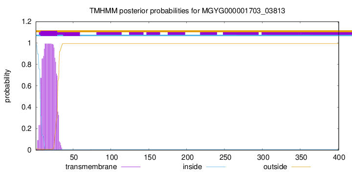
TMHMM annotations
Basic Information help
| Species | Vibrio fluvialis | |||||||||||
|---|---|---|---|---|---|---|---|---|---|---|---|---|
| Lineage | Bacteria; Proteobacteria; Gammaproteobacteria; Enterobacterales; Vibrionaceae; Vibrio; Vibrio fluvialis | |||||||||||
| CAZyme ID | MGYG000001703_03813 | |||||||||||
| CAZy Family | GH5 | |||||||||||
| CAZyme Description | Mannan endo-1,4-beta-mannosidase | |||||||||||
| CAZyme Property |
|
|||||||||||
| Genome Property |
|
|||||||||||
| Gene Location | Start: 32013; End: 33218 Strand: + | |||||||||||
CAZyme Signature Domains help
| Family | Start | End | Evalue | family coverage |
|---|---|---|---|---|
| GH5 | 32 | 282 | 1.5e-100 | 0.9881422924901185 |
CDD Domains download full data without filtering help
| Cdd ID | Domain | E-Value | qStart | qEnd | sStart | sEnd | Domain Description |
|---|---|---|---|---|---|---|---|
| pfam00150 | Cellulase | 2.78e-36 | 34 | 279 | 4 | 272 | Cellulase (glycosyl hydrolase family 5). |
| pfam02013 | CBM_10 | 1.46e-05 | 325 | 358 | 1 | 36 | Cellulose or protein binding domain. This domain is found in two distinct sets of proteins with different functions. Those found in aerobic bacteria bind cellulose (or other carbohydrates); but in anaerobic fungi they are protein binding domains, referred to as dockerin domains or docking domains. They are believed to be responsible for the assembly of a multiprotein cellulase/hemicellulase complex, similar to the cellulosome found in certain anaerobic bacteria. |
| pfam02013 | CBM_10 | 3.05e-05 | 363 | 394 | 3 | 36 | Cellulose or protein binding domain. This domain is found in two distinct sets of proteins with different functions. Those found in aerobic bacteria bind cellulose (or other carbohydrates); but in anaerobic fungi they are protein binding domains, referred to as dockerin domains or docking domains. They are believed to be responsible for the assembly of a multiprotein cellulase/hemicellulase complex, similar to the cellulosome found in certain anaerobic bacteria. |
| smart01064 | CBM_10 | 8.05e-05 | 363 | 394 | 3 | 29 | Cellulose or protein binding domain. This domain is found in two distinct sets of proteins with different functions. Those found in aerobic bacteria bind cellulose (or other carbohydrates); but in anaerobic fungi they are protein binding domains, referred to as dockerin domains or docking domains. They are believed to be responsible for the assembly of a multiprotein cellulase/hemicellulase complex, similar to the cellulosome found in certain anaerobic bacteria. |
| smart01064 | CBM_10 | 1.75e-04 | 326 | 358 | 2 | 29 | Cellulose or protein binding domain. This domain is found in two distinct sets of proteins with different functions. Those found in aerobic bacteria bind cellulose (or other carbohydrates); but in anaerobic fungi they are protein binding domains, referred to as dockerin domains or docking domains. They are believed to be responsible for the assembly of a multiprotein cellulase/hemicellulase complex, similar to the cellulosome found in certain anaerobic bacteria. |
CAZyme Hits help
| Hit ID | E-Value | Query Start | Query End | Hit Start | Hit End |
|---|---|---|---|---|---|
| AMF93023.1 | 5.89e-278 | 1 | 369 | 1 | 369 |
| QDC94577.1 | 2.80e-276 | 1 | 369 | 1 | 369 |
| ADT88758.1 | 9.35e-275 | 1 | 369 | 1 | 369 |
| BAA25188.1 | 1.49e-186 | 11 | 362 | 11 | 363 |
| AWB65998.1 | 5.76e-167 | 16 | 368 | 17 | 361 |
PDB Hits download full data without filtering help
| Hit ID | E-Value | Query Start | Query End | Hit Start | Hit End | Description |
|---|---|---|---|---|---|---|
| 1BQC_A | 3.56e-107 | 22 | 320 | 3 | 301 | Beta-MannanaseFrom Thermomonospora Fusca [Thermobifida fusca],2MAN_A Mannotriose Complex Of Thermomonospora Fusca Beta-Mannanase [Thermobifida fusca],3MAN_A Mannohexaose Complex Of Thermomonospora Fusca Beta-mannanase [Thermobifida fusca] |
| 4FK9_A | 3.99e-99 | 21 | 320 | 22 | 318 | HighResolution Structure of the Catalytic Domain of Mannanase SActE_2347 from Streptomyces sp. SirexAA-E [Streptomyces sp. SirexAA-E] |
| 1WKY_A | 1.13e-96 | 21 | 321 | 8 | 304 | Crystalstructure of alkaline mannanase from Bacillus sp. strain JAMB-602: catalytic domain and its Carbohydrate Binding Module [Bacillus sp. JAMB-602] |
| 3WSU_A | 9.99e-94 | 22 | 320 | 38 | 333 | Crystalstructure of beta-mannanase from Streptomyces thermolilacinus [Streptomyces thermolilacinus],3WSU_B Crystal structure of beta-mannanase from Streptomyces thermolilacinus [Streptomyces thermolilacinus] |
| 4Y7E_A | 8.00e-93 | 22 | 320 | 38 | 333 | Crystalstructure of beta-mannanase from Streptomyces thermolilacinus with mannohexaose [Streptomyces thermolilacinus],4Y7E_B Crystal structure of beta-mannanase from Streptomyces thermolilacinus with mannohexaose [Streptomyces thermolilacinus] |
Swiss-Prot Hits download full data without filtering help
| Hit ID | E-Value | Query Start | Query End | Hit Start | Hit End | Description |
|---|---|---|---|---|---|---|
| B3PF24 | 2.91e-132 | 21 | 369 | 47 | 399 | Mannan endo-1,4-beta-mannosidase OS=Cellvibrio japonicus (strain Ueda107) OX=498211 GN=man5A PE=1 SV=1 |
| P51529 | 7.90e-113 | 22 | 361 | 39 | 379 | Mannan endo-1,4-beta-mannosidase OS=Streptomyces lividans OX=1916 GN=manA PE=1 SV=2 |
| P22533 | 7.42e-90 | 42 | 313 | 55 | 326 | Beta-mannanase/endoglucanase A OS=Caldicellulosiruptor saccharolyticus OX=44001 GN=manA PE=1 SV=2 |
| G1K3N4 | 1.51e-88 | 21 | 321 | 1 | 297 | Mannan endo-1,4-beta-mannosidase OS=Salipaludibacillus agaradhaerens OX=76935 PE=1 SV=1 |
SignalP and Lipop Annotations help
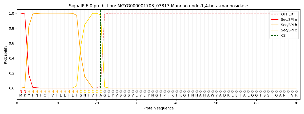
This protein is predicted as SP

| Other | SP_Sec_SPI | LIPO_Sec_SPII | TAT_Tat_SPI | TATLIP_Sec_SPII | PILIN_Sec_SPIII |
|---|---|---|---|---|---|
| 0.000349 | 0.998916 | 0.000211 | 0.000157 | 0.000163 | 0.000167 |

