You are browsing environment: HUMAN GUT
CAZyme Information: MGYG000001988_01387
You are here: Home > Sequence: MGYG000001988_01387
Basic Information |
Genomic context |
Full Sequence |
Enzyme annotations |
CAZy signature domains |
CDD domains |
CAZyme hits |
PDB hits |
Swiss-Prot hits |
SignalP and Lipop annotations |
TMHMM annotations
Basic Information help
| Species | Marvinbryantia sp900550755 | |||||||||||
|---|---|---|---|---|---|---|---|---|---|---|---|---|
| Lineage | Bacteria; Firmicutes_A; Clostridia; Lachnospirales; Lachnospiraceae; Marvinbryantia; Marvinbryantia sp900550755 | |||||||||||
| CAZyme ID | MGYG000001988_01387 | |||||||||||
| CAZy Family | GH0 | |||||||||||
| CAZyme Description | hypothetical protein | |||||||||||
| CAZyme Property |
|
|||||||||||
| Genome Property |
|
|||||||||||
| Gene Location | Start: 19051; End: 20316 Strand: + | |||||||||||
CDD Domains download full data without filtering help
| Cdd ID | Domain | E-Value | qStart | qEnd | sStart | sEnd | Domain Description |
|---|---|---|---|---|---|---|---|
| pfam01471 | PG_binding_1 | 1.05e-15 | 345 | 406 | 1 | 57 | Putative peptidoglycan binding domain. This domain is composed of three alpha helices. This domain is found at the N or C-terminus of a variety of enzymes involved in bacterial cell wall degradation. This domain may have a general peptidoglycan binding function. This family is found N-terminal to the catalytic domain of matrixins. The domain is found to bind peptidoglycan experimentally. |
| COG3409 | PGRP | 1.18e-10 | 322 | 404 | 103 | 181 | Peptidoglycan-binding (PGRP) domain of peptidoglycan hydrolases [Cell wall/membrane/envelope biogenesis]. |
| COG3409 | PGRP | 1.45e-08 | 340 | 407 | 39 | 103 | Peptidoglycan-binding (PGRP) domain of peptidoglycan hydrolases [Cell wall/membrane/envelope biogenesis]. |
| TIGR02669 | SpoIID_LytB | 9.66e-06 | 189 | 316 | 117 | 260 | SpoIID/LytB domain. This model describes a domain found typically in two or three proteins per genome in Cyanobacteria and Firmicutes, and sporadically in other genomes. One member is SpoIID of Bacillus subtilis. Another in B. subtilis is the C-terminal half of LytB, encoded immediately upstream of an amidase, the autolysin LytC, to which its N-terminus is homologous. Gene neighborhoods are not well conserved for members of this family, as many, such as SpoIID, are monocistronic. One early modelling-based study suggests a DNA-binding role for SpoIID, but the function of this domain is unknown. [Unknown function, General] |
| COG2385 | SpoIID | 0.001 | 293 | 316 | 362 | 385 | Peptidoglycan hydrolase (amidase) enhancer domain [Cell wall/membrane/envelope biogenesis]. |
CAZyme Hits help
| Hit ID | E-Value | Query Start | Query End | Hit Start | Hit End |
|---|---|---|---|---|---|
| AWY98201.1 | 4.92e-206 | 11 | 419 | 12 | 420 |
| QEI32542.1 | 1.06e-202 | 7 | 419 | 15 | 429 |
| QHB25032.1 | 1.06e-202 | 7 | 419 | 15 | 429 |
| QJU14725.1 | 1.09e-202 | 1 | 419 | 1 | 420 |
| QQQ92685.1 | 1.09e-202 | 1 | 419 | 1 | 420 |
PDB Hits download full data without filtering help
| Hit ID | E-Value | Query Start | Query End | Hit Start | Hit End | Description |
|---|---|---|---|---|---|---|
| 1LBU_A | 1.39e-07 | 343 | 408 | 13 | 78 | HydrolaseMetallo (zn) Dd-peptidase [Streptomyces albus G] |
| 7RUM_A | 2.63e-07 | 340 | 408 | 25 | 87 | ChainA, Endolysin [Salmonella phage GEC_vB_GOT],7RUM_B Chain B, Endolysin [Salmonella phage GEC_vB_GOT] |
| 5NM7_A | 3.20e-06 | 340 | 403 | 5 | 62 | Crystalstructure of Burkholderia AP3 phage endolysin [Burkholderia],5NM7_G Crystal structure of Burkholderia AP3 phage endolysin [Burkholderia] |
Swiss-Prot Hits download full data without filtering help
| Hit ID | E-Value | Query Start | Query End | Hit Start | Hit End | Description |
|---|---|---|---|---|---|---|
| P00733 | 8.66e-07 | 323 | 408 | 34 | 120 | Zinc D-Ala-D-Ala carboxypeptidase OS=Streptomyces albus G OX=1962 PE=1 SV=2 |
SignalP and Lipop Annotations help
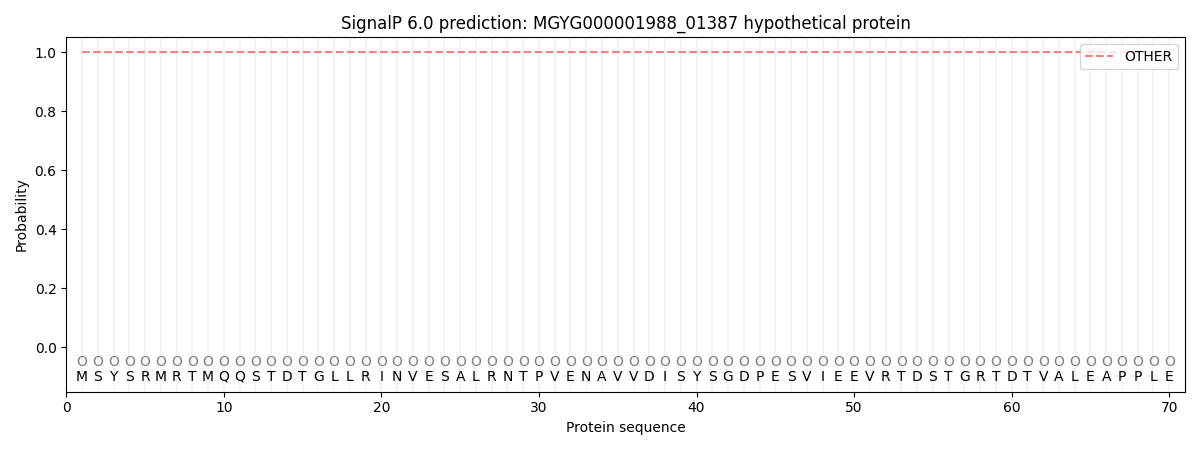
This protein is predicted as OTHER

| Other | SP_Sec_SPI | LIPO_Sec_SPII | TAT_Tat_SPI | TATLIP_Sec_SPII | PILIN_Sec_SPIII |
|---|---|---|---|---|---|
| 1.000041 | 0.000002 | 0.000000 | 0.000000 | 0.000000 | 0.000000 |
