You are browsing environment: HUMAN GUT
CAZyme Information: MGYG000002402_00572
You are here: Home > Sequence: MGYG000002402_00572
Basic Information |
Genomic context |
Full Sequence |
Enzyme annotations |
CAZy signature domains |
CDD domains |
CAZyme hits |
PDB hits |
Swiss-Prot hits |
SignalP and Lipop annotations |
TMHMM annotations
Basic Information help
| Species | Clostridium_H massiliodielmoense | |||||||||||
|---|---|---|---|---|---|---|---|---|---|---|---|---|
| Lineage | Bacteria; Firmicutes_A; Clostridia; Clostridiales; Clostridiaceae; Clostridium_H; Clostridium_H massiliodielmoense | |||||||||||
| CAZyme ID | MGYG000002402_00572 | |||||||||||
| CAZy Family | GH73 | |||||||||||
| CAZyme Description | hypothetical protein | |||||||||||
| CAZyme Property |
|
|||||||||||
| Genome Property |
|
|||||||||||
| Gene Location | Start: 154973; End: 156121 Strand: - | |||||||||||
CDD Domains download full data without filtering help
| Cdd ID | Domain | E-Value | qStart | qEnd | sStart | sEnd | Domain Description |
|---|---|---|---|---|---|---|---|
| COG1705 | FlgJ | 2.83e-52 | 2 | 152 | 41 | 188 | Flagellum-specific peptidoglycan hydrolase FlgJ [Cell wall/membrane/envelope biogenesis, Cell motility]. |
| NF038016 | sporang_Gsm | 2.15e-29 | 7 | 152 | 163 | 312 | sporangiospore maturation cell wall hydrolase GsmA. The peptidoglycan-hydrolyzing enzyme GsmA occurs in some sporangia-forming members of the Actinobacteria, such as Actinoplanes missouriensis, and is required for proper separation of spores. GsmA proteins have one or two SH3 domains N-terminal to the hydrolase domain. |
| pfam01832 | Glucosaminidase | 3.03e-29 | 12 | 97 | 1 | 77 | Mannosyl-glycoprotein endo-beta-N-acetylglucosaminidase. This family includes Mannosyl-glycoprotein endo-beta-N-acetylglucosaminidase EC:3.2.1.96. As well as the flageller protein J that has been shown to hydrolyze peptidoglycan. |
| cd06583 | PGRP | 1.91e-28 | 176 | 295 | 1 | 126 | Peptidoglycan recognition proteins (PGRPs) are pattern recognition receptors that bind, and in certain cases, hydrolyze peptidoglycans (PGNs) of bacterial cell walls. PGRPs have been divided into three classes: short PGRPs (PGRP-S), that are small (20 kDa) extracellular proteins; intermediate PGRPs (PGRP-I) that are 40-45 kDa and are predicted to be transmembrane proteins; and long PGRPs (PGRP-L), up to 90 kDa, which may be either intracellular or transmembrane. Several structures of PGRPs are known in insects and mammals, some bound with substrates like Muramyl Tripeptide (MTP) or Tracheal Cytotoxin (TCT). The substrate binding site is conserved in PGRP-LCx, PGRP-LE, and PGRP-Ialpha proteins. This family includes Zn-dependent N-Acetylmuramoyl-L-alanine Amidase, EC:3.5.1.28. This enzyme cleaves the amide bond between N-acetylmuramoyl and L-amino acids, preferentially D-lactyl-L-Ala, in bacterial cell walls. The structure for the bacteriophage T7 lysozyme shows that two of the conserved histidines and a cysteine are zinc binding residues. Site-directed mutagenesis of T7 lysozyme indicates that two conserved residues, a Tyr and a Lys, are important for amidase activity. |
| PRK05684 | flgJ | 2.43e-28 | 6 | 143 | 154 | 294 | flagellar assembly peptidoglycan hydrolase FlgJ. |
CAZyme Hits help
| Hit ID | E-Value | Query Start | Query End | Hit Start | Hit End |
|---|---|---|---|---|---|
| QPW59454.1 | 7.19e-128 | 1 | 238 | 1 | 237 |
| AEB77625.1 | 2.68e-85 | 1 | 358 | 1 | 383 |
| ABK61021.1 | 3.62e-76 | 1 | 292 | 1 | 289 |
| QPW54608.1 | 7.05e-71 | 1 | 170 | 1 | 169 |
| QPW58692.1 | 6.66e-68 | 1 | 170 | 1 | 169 |
PDB Hits download full data without filtering help
| Hit ID | E-Value | Query Start | Query End | Hit Start | Hit End | Description |
|---|---|---|---|---|---|---|
| 7F5I_A | 7.43e-37 | 175 | 298 | 29 | 149 | ChainA, amidase [Clostridium perfringens str. 13] |
| 6SRT_A | 2.18e-31 | 160 | 302 | 24 | 163 | EndolysineN-acetylmuramoyl-L-alanine amidase LysCS from Clostridium intestinale URNW [Clostridium intestinale URNW] |
| 6SSC_A | 4.10e-31 | 160 | 302 | 47 | 186 | N-acetylmuramoyl-L-alanineamidase LysC from Clostridium intestinale URNW [Clostridium intestinale] |
| 3FI7_A | 4.40e-17 | 4 | 152 | 30 | 183 | CrystalStructure of the autolysin Auto (Lmo1076) from Listeria monocytogenes, catalytic domain [Listeria monocytogenes EGD-e] |
| 5DN5_A | 9.29e-17 | 23 | 143 | 22 | 145 | Structureof a C-terminally truncated glycoside hydrolase domain from Salmonella typhimurium FlgJ [Salmonella enterica subsp. enterica serovar Typhimurium str. LT2],5DN5_B Structure of a C-terminally truncated glycoside hydrolase domain from Salmonella typhimurium FlgJ [Salmonella enterica subsp. enterica serovar Typhimurium str. LT2],5DN5_C Structure of a C-terminally truncated glycoside hydrolase domain from Salmonella typhimurium FlgJ [Salmonella enterica subsp. enterica serovar Typhimurium str. LT2] |
Swiss-Prot Hits download full data without filtering help
| Hit ID | E-Value | Query Start | Query End | Hit Start | Hit End | Description |
|---|---|---|---|---|---|---|
| O32083 | 9.77e-21 | 1 | 168 | 38 | 214 | Exo-glucosaminidase LytG OS=Bacillus subtilis (strain 168) OX=224308 GN=lytG PE=1 SV=1 |
| P37710 | 1.12e-20 | 5 | 243 | 181 | 427 | Autolysin OS=Enterococcus faecalis (strain ATCC 700802 / V583) OX=226185 GN=EF_0799 PE=1 SV=2 |
| P39046 | 2.67e-19 | 24 | 152 | 82 | 214 | Muramidase-2 OS=Enterococcus hirae (strain ATCC 9790 / DSM 20160 / JCM 8729 / LMG 6399 / NBRC 3181 / NCIMB 6459 / NCDO 1258 / NCTC 12367 / WDCM 00089 / R) OX=768486 GN=EHR_05900 PE=1 SV=1 |
| Q9V4X2 | 2.68e-17 | 181 | 293 | 48 | 169 | Peptidoglycan-recognition protein SC2 OS=Drosophila melanogaster OX=7227 GN=PGRP-SC2 PE=2 SV=1 |
| Q70PU1 | 3.69e-17 | 181 | 293 | 48 | 169 | Peptidoglycan-recognition protein SC2 OS=Drosophila simulans OX=7240 GN=PGRP-SC2 PE=3 SV=1 |
SignalP and Lipop Annotations help
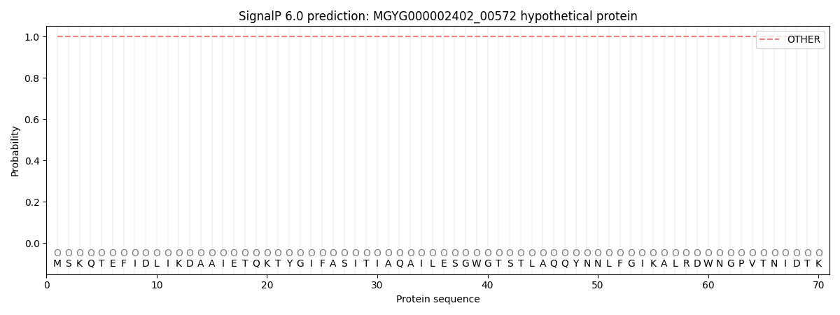
This protein is predicted as OTHER

| Other | SP_Sec_SPI | LIPO_Sec_SPII | TAT_Tat_SPI | TATLIP_Sec_SPII | PILIN_Sec_SPIII |
|---|---|---|---|---|---|
| 1.000010 | 0.000026 | 0.000000 | 0.000000 | 0.000000 | 0.000000 |
