You are browsing environment: HUMAN GUT
CAZyme Information: MGYG000002532_03510
You are here: Home > Sequence: MGYG000002532_03510
Basic Information |
Genomic context |
Full Sequence |
Enzyme annotations |
CAZy signature domains |
CDD domains |
CAZyme hits |
PDB hits |
Swiss-Prot hits |
SignalP and Lipop annotations |
TMHMM annotations
Basic Information help
| Species | Enterovibrio hollisae | |||||||||||
|---|---|---|---|---|---|---|---|---|---|---|---|---|
| Lineage | Bacteria; Proteobacteria; Gammaproteobacteria; Enterobacterales; Vibrionaceae; Enterovibrio; Enterovibrio hollisae | |||||||||||
| CAZyme ID | MGYG000002532_03510 | |||||||||||
| CAZy Family | CE4 | |||||||||||
| CAZyme Description | 2,3,4,5-tetrahydropyridine-2,6-dicarboxylate N-acetyltransferase | |||||||||||
| CAZyme Property |
|
|||||||||||
| Genome Property |
|
|||||||||||
| Gene Location | Start: 3823169; End: 3825508 Strand: + | |||||||||||
CAZyme Signature Domains help
| Family | Start | End | Evalue | family coverage |
|---|---|---|---|---|
| CE4 | 405 | 546 | 9.8e-35 | 0.9384615384615385 |
CDD Domains download full data without filtering help
| Cdd ID | Domain | E-Value | qStart | qEnd | sStart | sEnd | Domain Description |
|---|---|---|---|---|---|---|---|
| cd03819 | GT4_WavL-like | 4.22e-67 | 3 | 273 | 1 | 298 | Vibrio cholerae WavL and similar sequences. This family is most closely related to the GT4 family of glycosyltransferases. WavL in Vibrio cholerae has been shown to be involved in the biosynthesis of the lipopolysaccharide core. |
| cd10918 | CE4_NodB_like_5s_6s | 2.51e-60 | 409 | 568 | 1 | 157 | Putative catalytic NodB homology domain of PgaB, IcaB, and similar proteins which consist of a deformed (beta/alpha)8 barrel fold with 5- or 6-strands. This family belongs to the large and functionally diverse carbohydrate esterase 4 (CE4) superfamily, whose members show strong sequence similarity with some variability due to their distinct carbohydrate substrates. It includes bacterial poly-beta-1,6-N-acetyl-D-glucosamine N-deacetylase PgaB, hemin storage system HmsF protein in gram-negative species, intercellular adhesion proteins IcaB, and many uncharacterized prokaryotic polysaccharide deacetylases. It also includes a putative polysaccharide deacetylase YxkH encoded by the Bacillus subtilis yxkH gene, which is one of six polysaccharide deacetylase gene homologs present in the Bacillus subtilis genome. Sequence comparison shows all family members contain a conserved domain similar to the catalytic NodB homology domain of rhizobial NodB-like proteins, which consists of a deformed (beta/alpha)8 barrel fold with 6 or 7 strands. However, in this family, most proteins have 5 strands and some have 6 strands. Moreover, long insertions are found in many family members, whose function remains unknown. |
| cd10969 | CE4_Ecf1_like_5s | 4.87e-43 | 374 | 565 | 4 | 202 | Putative catalytic NodB homology domain of a hypothetical protein Ecf1 from Escherichia coli and similar proteins. This family contains a hypothetical protein Ecf1 from Escherichia coli and its prokaryotic homologs. Although their biochemical properties remain to be determined, members in this family contain a conserved domain with a 5-stranded beta/alpha barrel, which is similar to the catalytic NodB homology domain of rhizobial NodB-like proteins, belonging to the larger carbohydrate esterase 4 (CE4) superfamily. |
| cd04647 | LbH_MAT_like | 3.84e-38 | 658 | 767 | 3 | 109 | Maltose O-acyltransferase (MAT)-like: This family is composed of maltose O-acetyltransferase, galactoside O-acetyltransferase (GAT), xenobiotic acyltransferase (XAT) and similar proteins. MAT and GAT catalyze the CoA-dependent acetylation of the 6-hydroxyl group of their respective sugar substrates. MAT acetylates maltose and glucose exclusively while GAT specifically acetylates galactopyranosides. XAT catalyzes the CoA-dependent acetylation of a variety of hydroxyl-bearing acceptors such as chloramphenicol and streptogramin, among others. XATs are implicated in inactivating xenobiotics leading to xenobiotic resistance in patients. Members of this family contain a a left-handed parallel beta-helix (LbH) domain with at least 5 turns, each containing three imperfect tandem repeats of a hexapeptide repeat motif (X-[STAV]-X-[LIV]-[GAED]-X). They are trimeric in their active form. |
| cd10966 | CE4_yadE_5s | 1.05e-32 | 407 | 570 | 2 | 160 | Putative catalytic polysaccharide deacetylase domain of uncharacterized protein yadE and similar proteins. This family contains an uncharacterized protein yadE from Escherichia coli and its bacterial homologs. Although its molecular function remains unknown, yadE shows high sequence similarity with the catalytic NodB homology domain of outer membrane lipoprotein PgaB and the surface-attached protein intercellular adhesion protein IcaB. Both PgaB and IcaB are essential in bacterial biofilm formation. |
CAZyme Hits help
| Hit ID | E-Value | Query Start | Query End | Hit Start | Hit End |
|---|---|---|---|---|---|
| AMG30447.1 | 0.0 | 1 | 779 | 1 | 779 |
| AYV21232.1 | 8.31e-300 | 1 | 587 | 1 | 587 |
| QJY37746.1 | 2.88e-296 | 1 | 593 | 1 | 590 |
| QPG36570.1 | 2.88e-296 | 1 | 593 | 1 | 590 |
| ANJ24059.1 | 1.04e-295 | 1 | 586 | 1 | 586 |
PDB Hits download full data without filtering help
| Hit ID | E-Value | Query Start | Query End | Hit Start | Hit End | Description |
|---|---|---|---|---|---|---|
| 6GO1_A | 1.53e-19 | 342 | 571 | 95 | 310 | CrystalStructure of a Bacillus anthracis peptidoglycan deacetylase [Bacillus anthracis],6GO1_B Crystal Structure of a Bacillus anthracis peptidoglycan deacetylase [Bacillus anthracis] |
| 4V33_A | 1.19e-18 | 342 | 578 | 139 | 359 | Crystalstructure of the putative polysaccharide deacetylase BA0330 from bacillus anthracis [Bacillus anthracis],4V33_B Crystal structure of the putative polysaccharide deacetylase BA0330 from bacillus anthracis [Bacillus anthracis] |
| 6DQ3_A | 2.16e-18 | 347 | 574 | 9 | 224 | ChainA, Polysaccharide deacetylase [Streptococcus pyogenes],6DQ3_B Chain B, Polysaccharide deacetylase [Streptococcus pyogenes] |
| 4HD5_A | 9.31e-18 | 340 | 578 | 137 | 359 | CrystalStructure of BC0361, a polysaccharide deacetylase from Bacillus cereus [Bacillus cereus ATCC 14579] |
| 3JQY_A | 7.69e-16 | 641 | 773 | 92 | 220 | CrystalStructure of the polySia specific acetyltransferase NeuO [Escherichia coli],3JQY_B Crystal Structure of the polySia specific acetyltransferase NeuO [Escherichia coli],3JQY_C Crystal Structure of the polySia specific acetyltransferase NeuO [Escherichia coli] |
Swiss-Prot Hits download full data without filtering help
| Hit ID | E-Value | Query Start | Query End | Hit Start | Hit End | Description |
|---|---|---|---|---|---|---|
| P94361 | 5.29e-25 | 343 | 568 | 61 | 265 | Putative polysaccharide deacetylase YxkH OS=Bacillus subtilis (strain 168) OX=224308 GN=yxkH PE=3 SV=1 |
| A1ADJ6 | 9.72e-15 | 641 | 773 | 155 | 283 | Polysialic acid O-acetyltransferase OS=Escherichia coli O1:K1 / APEC OX=405955 GN=neuO PE=1 SV=1 |
| P37515 | 1.93e-11 | 655 | 768 | 93 | 182 | Probable maltose O-acetyltransferase OS=Bacillus subtilis (strain 168) OX=224308 GN=maa PE=3 SV=1 |
| Q86A05 | 2.79e-10 | 687 | 771 | 108 | 193 | Putative acetyltransferase DDB_G0275507 OS=Dictyostelium discoideum OX=44689 GN=DDB_G0275507 PE=3 SV=1 |
| P23364 | 8.70e-10 | 637 | 768 | 40 | 161 | Chloramphenicol acetyltransferase OS=Agrobacterium fabrum (strain C58 / ATCC 33970) OX=176299 GN=cat PE=3 SV=1 |
SignalP and Lipop Annotations help
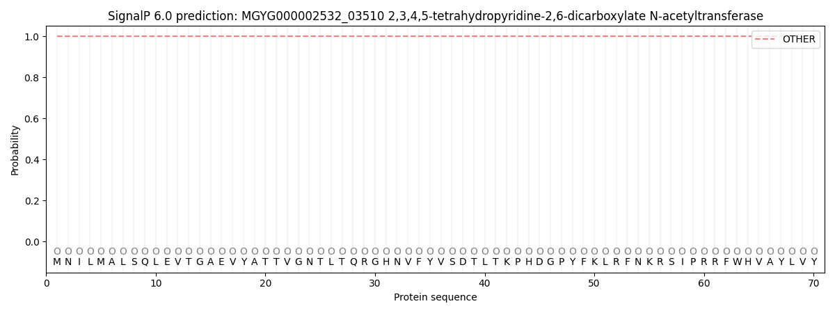
This protein is predicted as OTHER

| Other | SP_Sec_SPI | LIPO_Sec_SPII | TAT_Tat_SPI | TATLIP_Sec_SPII | PILIN_Sec_SPIII |
|---|---|---|---|---|---|
| 0.999836 | 0.000158 | 0.000013 | 0.000000 | 0.000000 | 0.000004 |
